X-ray MHz tomoscopy – state of the art
There are few alternative ways how to achieve the 3D imaging of rapidly evolving samples and objects of all kinds, but nothing compares directly to X-ray MHz tomoscopy. The reason is in unique imaging potential of the latest coherent X-ray sources, enabling advanced experimental setups, combined with new data acquisition and data reconstruction processes.
Time-frames
As in conventional photography, the object pictures are sharp – “frozen” in time, when object illumination – flash strobe – lasts much shorter time than the time to move object or its part significantly.
In our case, the X-ray photon sources provide sequence of very short and bright flashes. For our most capable source – at EuXFEL [facility overview] – the pulse is lasting typically only 30 fs, although they can be made to last shorter (up to 10 fs) or longer (say 100 fs) times [parameters]. In 30 fs even light passes just about 10 µm in vacuum.
This is the upper limit defining motion blur – most of the processes happen at speeds orders and orders of magnitude lower than the speed of light.
The pulse repetition rate is programmable, in EuXFEL for now up to 220 ns period between the individual pulses. This allows, for short time, to watch time evolution of the sample at “frozen moments” in time.
Opacity
By using X-rays with very short wavelength, the photons can penetrate any material to great depth before significant attenuation due to photon absorption occurs. The potential to “see through the matter” was clearly recognised from the very beginning and awarded by the very first Nobel prize to W. C. Röntgen. Hard X-ray imaging allows to see things, otherwise unobservable.
Coherence and phase contrast
X-ray free electron lasers serve as extremely brilliant source of X-ray photons with very high monochromaticity and coherence, which allows phase contrast imaging. Similarly to well-known optical microscopy, phase contrast provides superior image to absorption contrast, especially for objects, optical properties of which differ very little from those of surrounding.
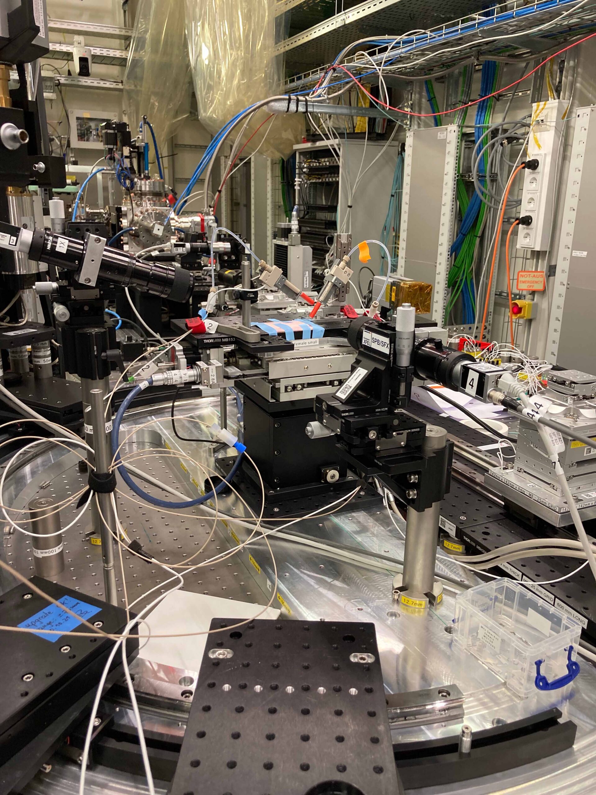
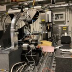
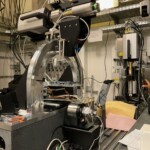
The beginning of the setup at SPB/SFX beamline. In front, part of the microfluidic device for liquid sample delivery and calibration. June 2022

